Allegation:
A high-quality visual representation accurately depicts a 1 cubic millimeter sample of the human brain.
Evaluation:

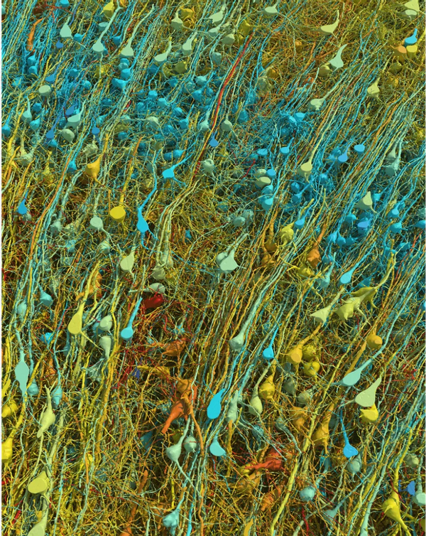
With thin stripes of blue, green and orange bursting out on a black background, image Posted on Reddit on May 12, 2024, it claimed to show “1 cubic millimeter of brain.” At the time of this writing, the post had received more than 26,000 upvotes.
A. Google keyword search returned dozens of relevant results, including Smithsonian Magazine The article, published in May 2024, describes researchers who created “a digital map showing a small part of the human brain in unprecedented detail.”
This image is original and actually shows a 3D map of neurons in the brain. We rated this claim as “True”, but first let’s explain what exactly this research means and what stands out about the visualization.
The Smithsonian article cited an article published by . Nature News outlined this research. Scientists at Google Research and Harvard University’s Lichtman Laboratory have mapped a small part of the human brain, the details of which have been published in a peer-reviewed journal. Science. The images show neurons, or connections, between brain cells responsible for sharing information throughout the organ.
The brain sample was taken from the cortex, the part of the brain involved in learning, from a 45-year-old woman who had surgery to treat epilepsy. The sample, stained and preserved with heavy metals to make it more visible, was cut into an estimated 5,000 slices just 34 nanometers thick to be viewed using electron microscopes.
According to the Science article, the 3D brain map:
… it occupies a volume of about one cubic millimeter, or one millionth of the entire brain, and contains roughly 57,000 cells and 150 million synapses, or connections between neurons… [and] It contains a massive 1.4 petabytes of data.
AI models custom-built by the researchers were used to stitch together the images to reconstruct the sample in three dimensions with relevant datasets and visualizations. made available online.
The “reconstruction of a millimeter-scale piece of the human cerebral cortex at nanoscale resolution” is said to provide an unprecedented view of how brain tissue is organized in various layers, including supracellular, cellular and subcellular. newsletter In that case.
The reconstruction contains an estimated “57,000 cells, approximately 230 millimeters of blood vessels, and approximately 150 million synapses,” according to the authors. Thanks to these, the research team discovered previously underappreciated aspects of the human temporal cortex.
Disruption of synaptic and neural brain circuits that convey information for brain functionality is thought to play a role in a number of disorders rooted in the brain. schizophrenia and autism with bipolar disorder and the other neuropsychiatric diseases. By better capturing how these communication pathways work, scientists hope to gain a more insightful understanding of how such states form and persist in the brain.
For the study at hand, the researchers concluded that this provided evidence for a new approach to “visualize and ultimately gain insight into the physical underpinnings of normal and disordered human brain function.”
The image used in the Reddit post is also a blog post It was published by Google on May 4, 2024, and explained how the image and others were created with the help of artificial intelligence. It read in part:
By combining brain imaging with AI-based image processing and analysis, our teams reconstructed nearly every cell and all their connections in a small volume of human brain tissue about half the size of a grain of rice.
Other images included in the study showed a “dense and complex map” of some pairs of neurons that had “the astonishing property of being extremely strongly connected to each other – through up to 50 synapses.”
(Google Search and Lichtman Lab (Harvard University). Renderings: D. Berger (Harvard University))
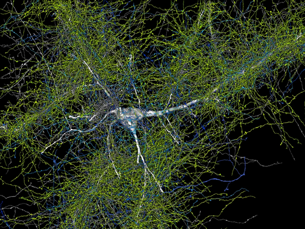
Another close-up showed synapses “swimming” between neurons, bringing with them information and signals essential to human life.
(Google Search and Lichtman Lab (Harvard University). Renderings: D. Berger (Harvard University))
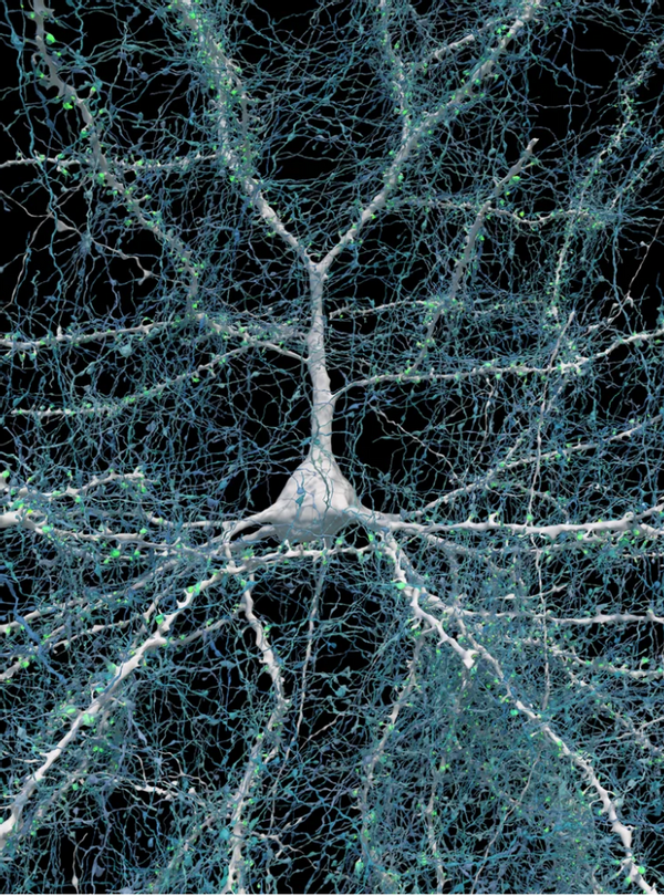
This reception of signals is illustrated in the image below, which shows a single neuron in white with its surrounding axons, a part of the neuron that sends electrical signals.
(Google Search and Lichtman Lab (Harvard University). Renderings: D. Berger (Harvard University))
“There is still much to observe and understand in the reconstruction of this part of the human brain, and we hope that other researchers will use the data to make additional discoveries.” Wrote Google team.
“Scientists believe that by continuing research into the connections in the brain, they may one day understand things like how our memories are formed or what causes neurological disorders and diseases such as autism and Alzheimer’s.”
Snopes examined other claims as well cerebral“From whether this is true to whether Bluetooth headsets exist, we only use 10% of our gray matter.”fry our brains.” Check out our other health rebuttals Here.
Resources:
“6 Incredible Images of the Human Brain Created with the Help of Google’s Artificial Intelligence.” GoogleMay 9, 2024, https://blog.google/technology/research/google-ai-research-new-images-human-brain/.
Anonymous. Making and Breaking Connections in the Brain | UC Davis Neuroscience Center. September 11, 2020, https://neuroscience.ucdavis.edu/news/making-and-breaking-connections-brain.
“Cubic Millimeter Piece of Human Brain Reconstructed at Nanoscale Resolution.” EurekAlert!, https://www.eurekalert.org/news-releases/1043546. Access date: 23 May 2024.
Magazine, Smithsonian and Will Sullivan. “Scientists Imaged and Mapped a Small Piece of the Human Brain. Here’s What They Found.” Smithsonian Magazine, https://www.smithsonianmag.com/smart-news/scientists-imaged-and-mapped-a-tiny-piece-of-human-brain-heres-what-they-found-180984340/. Access date: 23 May 2024.
Photo of 1 Cubic Millimeter Brain – Google Search. https://www.google.com/search?q=photograph+of+1+cubic+millimeter+brain&sca_esv=5a8c8ac8ff46dd80&sca_upv=1&ei=jJ1PZouQE-eA0PEP7NeKkAI&ved=0ahUKEwiLhvmtxaSGAxVnADQIHeyrAiIQ4dUDCBE&uact=5&oq=photograph +1+cubic+millimeter+brain&gs_lp= Egxnd3Mtd2l6LXNlcnAiJnBob3RvZ3JhcGggb2YgMSBjdWJpYyBtaWxsaW1ldGVyIGJyYWluMggQABiABBiiBDIIEAAYgAQYogQyCBAAGIAEGKIEMggQABiABBIiBDIIEAAYgAQYog RIuwxQ2QdY2QdwA3 gAkAEAmAGBAaABgQGqAQMwLjG4AQPIAQD4AQGYAgSgAooBwgiIOEAAYgAQYsAMYhgMYigXCAgsQABiwAxiiBBiJBcICCxAAGIAEGLADGKIEmAMAiAYBkAYIkgcDMy4xoAfpAw&sclient=gws-wiz-serp. Access date: 23 May 2024.
Published Data | H01 Version. https://h01-release.storage.googleapis.com/data.html. Access date: 23 May 2024.
Shapson-Coe, Alexander et al. “Petavoxel Fragment of Human Cerebral Cortex Reconstructed at Nanoscale Resolution.” Science, vol. 384, no. 6696, May 2024, p. eadk4858. DOI.org (Cross reference)https://doi.org/10.1126/science.adk4858.
Taoufik, Era et al. “Synaptic Dysfunction in Neurodegenerative and Neurodevelopmental Diseases: Overview of Induced Pluripotent Stem Cell-Based Disease Models.” Open Biology, vol. 8, no. September 9, 2018, p. 180138. PubMed Centerhttps://doi.org/10.1098/rsob.180138.
Wang, Xinyuan et al. “Synaptic Dysfunction in Complex Psychiatric Disorders: From Genetics to Mechanisms.” Genome Medicine, vol. 10, no. January 1, 2018, p. 9. BioMed Centerhttps://doi.org/10.1186/s13073-018-0518-5.
Wong, Carissa. “Cubic Millimeters of the Brain Mapped in Extraordinary Detail.” Nature, vol. 629, no. 8013, May 2024, p. 739–40. www.nature.comhttps://doi.org/10.1038/d41586-024-01387-9.